Dansk  Webmaster  Search   


SIJ - normal anatomy
SIJ - normal appearance by MRI
SIJ - anatomical variations
SIJ - pitfalls by MRI
Lumbosacral transitional vertebra
Lumbosacral transitional vertebra is a common variation, occurring in up to 25% of the population. It can either encompass sacralization of the fifth lumbar vertebral body or lumbarization of the S1 segment. There is a wide variety of morphologies ranging from broadened transverse processes to complete fusion occurring bilaterally, symmetrically or asymmetrically as well as unilaterally, doi:10.1097/00007632-198407000-00014.
 

Drawing illustrating the different variants of transitional vertebra from the publication of Castellvi et al., 1984, doi:10.1097/00007632-198407000-00014.


There may be accompanying low back pain, especially if there is persisting cleavage between the transverse process and the upper border of the sacrum (Castellvi type I and type II). Concomitant BME, fat deposition and/or sclerosis may occur, usually confined to pseudo-articulations and as such only occasionally reaching the SIJ possibly causing confusion with axSpA sacroiliitis changes.

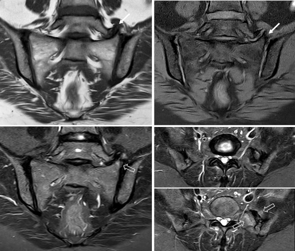
MRI in a 41-year-old woman with recurrent low back pain during several years, often with inflammatory characteristics; semi-coronal T1, T1FS and STIR image in addition to two semi-axial STIR images at the level of the transitional vertebra. There is enlargement of the left transverse process of L5 articulating with the upper border of the sacrum (Castellvi type IIA) accompanied by slight subchondral BME (open arrow) and also subchondral sclerosis and osteophyte formation (arrows).

Supplementary radiograph confirmed the finding and delineate the structural changes more clearly.
 
Visitors since May 1st., 2008:  1767312