Dansk  Webmaster  Search   


SIJ - normal anatomy
SIJ - normal appearance by MRI
SIJ - anatomical variations
SIJ - pitfalls by MRI
Lumbosacral transitional vertebra
SIJ anatomical variations

Anatomical SIJ variations are frequent findings, especially in females and awareness of their presence is important when interpreting MRI of the SIJ as they may represent pitfalls.

Seven SIJ variations have been described: Accessory SIJ, iliosacral complex, bipartite iliac bony plate, semicircular defects, crescent-like iliac bony plate, dysmorphic SIJ and unfused nuclei at the sacral wing.

The detection and description of six SIJ variations have primarily been based on CT, but they are also detectable by MRI which in addition can visualize concomitant BME, sclerosis and/or fat deposition. The occurrence of dysmorphic SIJ has primarily been based on MRI findings, doi:10.1007/s00256-021-03843-3; doi:10.1007/s00330-018-5540-x.

Drawing illustrating the six SIJ variants described in two major CT analyses with an estimation of there prevalence in the general population (Demir et al. 2007 and Prassopoulos et al. 1999).

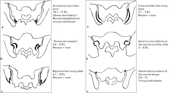
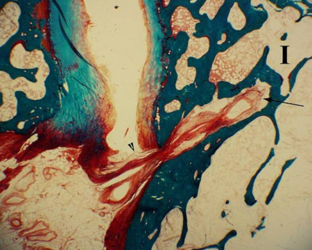
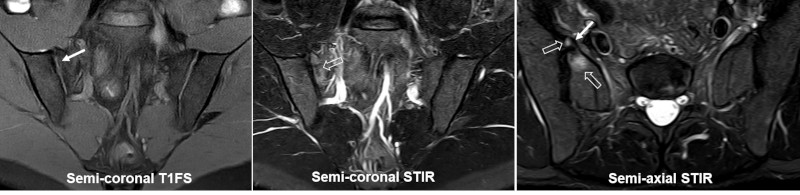
Only two of the variants described, dysmorphic SIJ and unfused nuclei, occur at the cartilaginous SIJ compartment and may therefore directly cause MRI changes, such as subchondral edema, simulating sacroiliitis confined to the cartilaginous joint compartment. BME adjacent to accessory SIJ and iliosacral complex will be located to the ligamentous joint compartment where BME areas will not be mistaken for sacroiliitis changes. However, the presence of variants in the ligamentous compartment may elicit strain-related changes in the cartilaginous joint compartment, often located anteriorly in the sacrum, doi:10.1007/s00256-021-03843-3, an area often presenting strain-related edema.

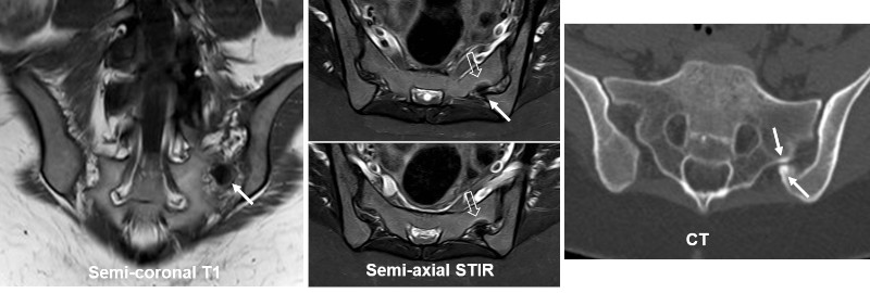
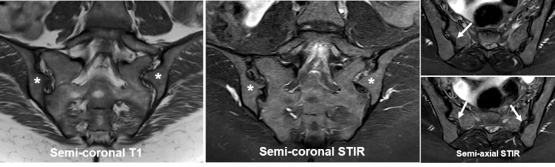
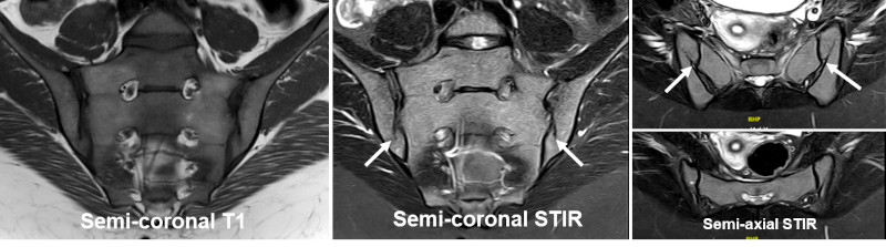
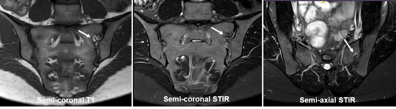
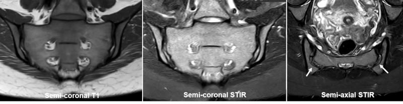
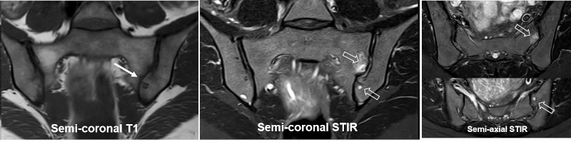
The MRI appearance of the different variations is shown beneath.

Accessory SIJ

Accessory (ACC) SIJ is an additional small joint in the ligamentous joint compartment. It can be uni- or bilateral and is mostly seen at the level of S2, but can also occur at S1 and S3, doi:10.1002/ca.20772. ACC SIJ is best visualized on semi-axial slices, but can be confirmed in the semi-coronal plane by the lack of interposed fat and ligaments between the joint facets.

MRI showing ACC SIJ (arrows) in a patient presenting with pain. There is concomitant subchondral sclerosis at the ACC joint (white arrows on T1) and a brim of osseous edema in the sacrum (open arrows). A concomitant semi-coronal CT reconstruction shown to the right clearly visualize the ACC SIJ.

 
Iliosacral complex

Iliosacral complex consists of an osseous protrusion of the iliac bone in the ligamentous joint compartment with a corresponding groove in the opposing sacral bone visualized in both the semi-coronal and semi-axial plane.


MRI showing bilateral iliosacral complex with iliac protrusion (asterisk) and a corresponding groove in the opposing sacral bone (arrows).

 
Bipartite iliac bony plate

Bipartite iliac bony plate is a cleft-like appearance posteriorly and inferiorly in the iliac bone best seen in the semi-axial plane. It is formed by a deep iliac insertion of an iliosacral ligament which is often surrounded by vessels, which can give rise to edematous changes, doi:10.1055/s-0034-1375574.

MRI showing bilateral bipartite iliac bony plate detectable on both the coronal and axial slices(arrows), but being most obvious on the axial slices .

For comparison, a histological section showing the ligament (arrowhead) inserting deep within the iliac bone.

 
Semicircular defects

A semicircular defect is a round well-defined defect in the iliac and/or sacral bone superiorly in the ligamentous joint compartment.

MRI showing a left-sided semicircular defect (arrows).

 
Crescent-like iliac bony plate

Crescent-like iliac bony plate is a concave configuration of the iliac bone posteriorly/superiorly in the ligamentous joint compartment with a congruent bulging of the opposite sacral bone.

MRI showing a bilateral crescent-like iliac bony plate with a concave configuration of the iliac bones (arrows).

 
Dysmorphic SIJ

Dysmorphic SIJ is a term used for abnormal osseous protrusions in the cartilaginous joint compartment, either of the sacrum or the ilium. It can be accompanied by strain-related subchondral edema and/or erosion-like osseous irregularities and thereby mimic sacroiliitis, doi:10.1007/s00256-021-03843-3; doi:10.1007/s00330-018-5540-x.


MRI in a patient with low back and buttock pain showing dysmorphic SIJ changes on the left side with the ileum protruding into the sacrum (arrow on T1). There is concomitant BME in the sacrum adjacent to the dysmorphic changes and also a subchondral cyst inferiorly in the ileum (open arrows), probably elicited by strain.

 
Unfused nuclei at the sacral wing

Nuclei at the upper sacral corners (sacral wing), which are normal developmental findings in childhood, may occasionally persist in adulthood and can be associated with adjacent BME.


MRI in a 26-year-old man with right-sided buttock pain showing persistence of a sacral nucleus (arrows) with accompanying BME both in the nucleus and in the sacral bone (open arrows).

 
Visitors since May 1st., 2008:  1765320