|
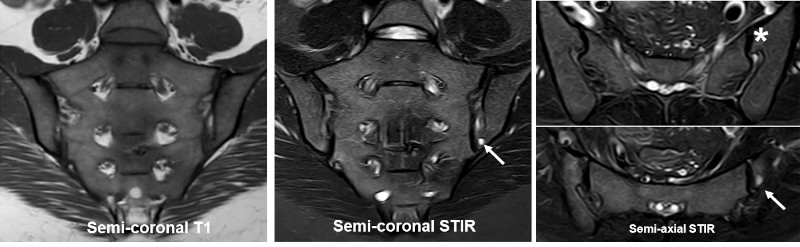
The frequent anatomical variations in the SIJ region, especially in females, may give rise to pitfalls. There can be concomitant BME and also erosion-like lesions simulating sacroiliitis. Although only two of the variants, dysmorphic SIJ and unfused nuclei, occur at the cartilaginous SIJ compartment where sacroiliitis changes are located, the presence of variants in the ligamentous compartment may elicit strain-related changes in the cartilaginous joint compartment, often located anteriorly in the sacrum. Besides, bipartite iliac bony plate can be misinterpreted as inflammatory changes. It is caused by a ligament attached deeply in the iliac bone at the border between the cartilaginous and the ligamentous joint portion. The ligaments are surrounded by vessels implying increased signal intensity on STIR images and may on coronal slices simulate erosion, doi:10.1055/s-0034-1375574. |