Dansk  Webmaster  Search   


SIJ - normal anatomy
SIJ - normal appearance by MRI
SIJ - anatomical variations
SIJ - pitfalls by MRI
Lumbosacral transitional vertebra
Normal SIJ anatomy macroscopically

The sacroiliac joints (SIJs) are built to be stable during the transmission of forces from the body to the lower extremities. This stability is obtained by obliquely orientated undulating joint facets, surrounding joint ligaments, and also strong external ligaments and muscles, doi:10.1111/j.1469-7580.2012.01564.x.

The images show the joint localization and stabilizing muscles in addition to some gender differences regarding load on the joints in the upright position.


The jSIJ is composed of two compartments: a ventral C-shaped cartilaginous compartment and a dorsal ligamentous compartment containing strong inter-osseous ligaments. The cartilaginous joint surfaces vary considerably in size, shape and contour, especially in females, and are usually smaller in females than in males, although the joint varies greatly in size, shape and contour. Due to an often backward tilted sacrum in females, the cartilaginous joint surface is usually more horizontally located and is smaller in females than in males. The SIJ in males is typically formed corresponding to the sacral segments S1, S2 and S3 whereas inclusion of the whole S3 segment is uncommon in females, doi:10.1111/j.1469-7580.2012.01564.x

3D CT-reconstructions of the sacral joint surface showing sex differences with a more horizontally located cartilaginous joint surface facet in woman than in man.

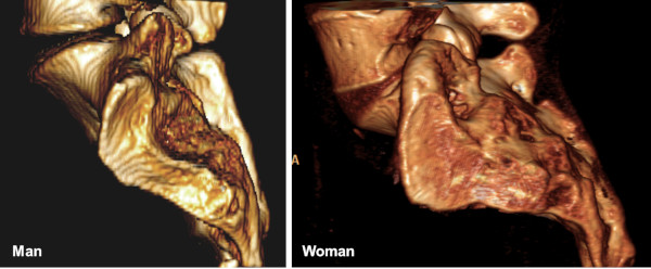
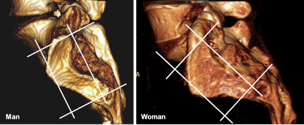
The 3D CT-reconstructions of the sacral joint surface in a woman and a man, respectively, with lines indicating the orientation of the two perpendicular slice orientations by MRI (semi-coronal and semi-axial) being located in relation to the upper vertebral plate of S5 or the posterior aspect of S2.


To obtain a clear delineation between the two different joint compartments axial visualization of the joint is needed showing the cartilaginous compartment ventrally and the ligamentous compartment posteriorly.

Images delineating the joint portions on an axial drawing and a axial histologic slice showing the two different joint compartments, a cartilaginous compartment ventrally (V) and the ligamentous compartment posteriorly (D). SC and IC = sacral and iliac cartilaginous; SL and IL = sacral and ligamentous.

 
Microscopic SIJ anatomy

The microscopic anatomy is rather complex. The cartilaginous compartment of the joint is without synovia (a symphysis) proximally whereas there are small synovial recesses at the distal part of the joint. The anatomy of the middle part is rather variable with a variety of normal variations giving raise to pitfalls at MR imaging, doi:10.1055/s-0034-1375574.


Microscopic section - proximal joint portion. In the proximal portion of the cartilaginous joint compartment there are strong ligaments connecting the joint facets anterior and proximally (VSIL = ventral sacroiliac ligament and PSIL = proximal sacroiliac ligament). Posteriorly the ligaments merge with the cartilage forming fibrocartilaginous-like tissue. There is no synovia in this region. I = ileum; S = sacrum; FC = fibrocartilage.

Microscopic section - distal joint portion. In the distal joint portion there are small recesses lined by synovia both anterior and posterior (arrows).

 
Visitors since May 1st., 2008:  1767797