Dansk  Webmaster  Search   


SIJ - normal anatomy
SIJ - normal appearance by MRI
SIJ - anatomical variations
SIJ - pitfalls by MRI
Lumbosacral transitional vertebra
Normal appearance of the SIJ by MRI

In the evaluation of the SIJ by MRI it is important to be aware of the normal joint appearance on semi-coronal and semi-axial slice orientation. The different joints portions are shown in the figure beneath.

MRI in a healthy young person, semi-coronal T1, T1FS and STIR image in addition to two semi-axial STIR images at the upper and lower part of the SIJ, respectively. The coronal slices located in the middle of the cartilaginous joint compartment visualize a normal joint space and subchondral bone without subchondral edema or fat deposition. The axial slices display both joint compartments with the cartilaginous joint portion located anteriorly (marked with # at the sacral side) and the ligamentous portion posteriorly (marked with asterisks at the iliac side of the joints). On these standard sequences the normal capsular structures may not be seen. Only the upper capsule on the right side is visible (arrow). This is partly due to the sequences used, but the capsule may also be invisible on dedicated high resolution images as shown beneath as they can be slim.


Semi-axial T1FS MR image not visualizing the ventral sacroiliac ligament on the left side, but it is clearly demonstrated on the right side (arrow). A microscopic image showing the slim capsule anteriorly is shown for comparison. 


The synovial pouches at the distal part of the joint may sometimes be visible in healthy persons.

STIR image demonstrating fluid filled pouches anteriorly as well as posteriorly at both SIJs (arrow). A microscopic image of the region anteriorly with an arrow on the synovial pouch is shown for comparison.

 
Bone marrow signals and structural changes

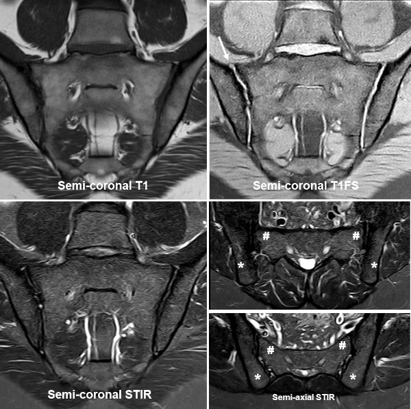
MR signal intensity in the bone marrow varies in healthy persons both on STIR/T2FS and T1-weighted sequences. Minor areas of increased subchondral signal on STIR/T2FS often occur in individuals above 30 years, but the extent is limited and deep and/or intense BME is rare, most frequently occurring anteriorly in the sacrum corresponding to the strain-related areas, doi:10.1002/art.42145.

On T1-weighted images there can be a patchy distribution of fat within the bone marrow with an increasing prevalence with increasing age and especially seen in a degenerative setting.

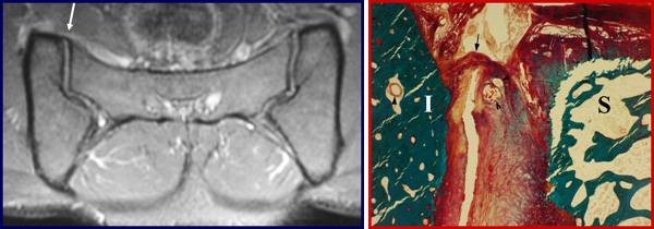
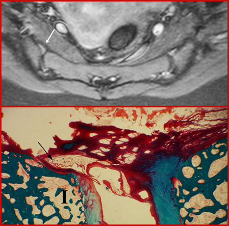
Erosion or erosion-like lesions may also occur as a normal finding, especially in middle aged and elderly persons. Besides, sclerosis, osteophytes and joint space alterations can occasionally be detected, but these features are best visualized by CT being detected with increasing frequency with age, doi:10.1016/S0009-9260(98)80316-4.

 
Visitors since May 1st., 2008:  1766377