|

|
Degenerative SIJ changes are frequent in middle-aged and older individuals, but rare below 30 years of age. It can occur with and without accompanying symptoms, doi:10.2106/JBJS.N.01101.
The imaging findings have mainly been described based on CT features consisting of joint-space narrowing, subchondral sclerosis and osteophyte formation in addition to the occasional occurrence of intraarticular air, subchondral cysts, ankylosis and sometimes small erosion-like lesions, but not manifest erosions as in axSpA. Developmental anomalies such as transitional vertebrae and accessory SI joints, predispose to degenerative changes, doi:10.1038/S41598-021-85303-5.
Unfortunately, degenerative changes can sometimes be difficult to detect with certainty by MRI where the exact joint space width can be difficult to estimate without appropriate cartilage sequences and small osteophytes may vanish due to the relatively thicker MR-slices compared to CT-slices. However, in addition to the changes best documented by CT, MRI may reveal dispersed fat deposition and/or subchondral BME usually occurring at the anterior superior/middle strain-related joint areas or at areas with osteophytes. However, BME is usually minimal, often visible on only one or two slices. In the case of such changes look for other signs suggestive of degenerative disease (joint space narrowing, subchondral sclerosis, osteophytes, joint vacuum phenomenon and subchondral cysts). |
|
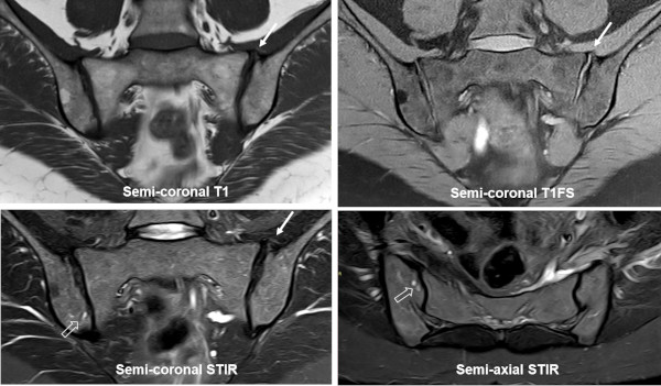
MRI in a 51-year-old woman complaining of low back pain, semi-coronal T1, T1FS and STIR, and semi-axial STIR image. There is osteophyte formation superiorly at the left SIJ (arrows) with concomitant slight iliac sclerosis. The joint spaces seem narrow caudally and there is concomitant subchondral cysts (open arrows), but no signs of erosions. |
|
|
|
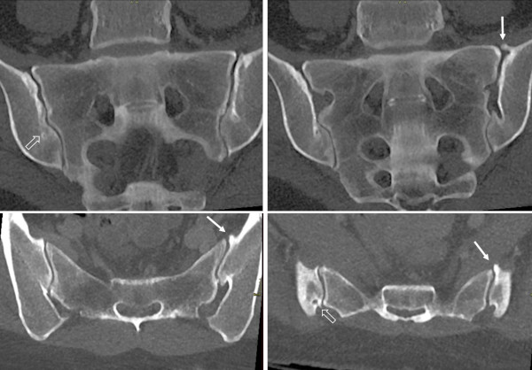
A CT performed some months previously, semi-coronal and semi-axial reconstructions, more clearly shows the osteophytes (arrows), the brim of iliac sclerosis and the subchondral cysts in addition to narrowed joint spaces caudally. |
|
|
| |
|
DISH is a common condition in individuals >50 years of age. It is a non-inflammatory condition characterized by pathologic calcification of entheses, primarily at the spine, but also at other joints, including the SIJ, doi:10.1007/S11926-020-00972-X. Changes are often asymptomatic, but patients may present with back pain and stiffness.
The imaging diagnosis of DISH is mainly based on flowing ossifications along the anterior border of the spine extending over >4 vertebral bodies. DISH changes at the SIJ similarly present as coarse bony/ossified bridges located to the SIJ ligaments and capsule at the upper part of the joints, which by radiography may resemble obliteration of the SIJ and simulate axSpA ankylosis. However, the lower two-thirds of the SIJ are usually spared, doi:10.2214/AJR.16.16994. Slight BME and fatty marrow metaplasia at the SIJ may occur, but erosions and subchondral sclerosis are rare, thus differentiating the changes from axSpA changes. |
|
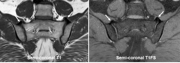
MRI in a 62-year-old man presenting with flowing thoracic paravertebral ossification conforming to DISH, semi-coronal T1 and T1FS of the SIJs showing new bone formation superiorly and anteriorly at the joints (arrows) in addition to a small area with subchondral fat deposition in the sacrum (open arrow), but no erosions and there was no BME. |
|
|
| |
|
|
|
|