Dansk  Webmaster  Search   


SIJ - differentials
OCI / postpartum changes
Strain-related SIJ changes
Degenerative / DISH changes
Infectious sacroiliitis
Fractures
Inflammatory conditions
Metabolic disorders
Tumors
Spine - differentials
Degenerative changes
Infectious spondylitis
Osteitis condensans ilii (OCI)

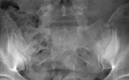
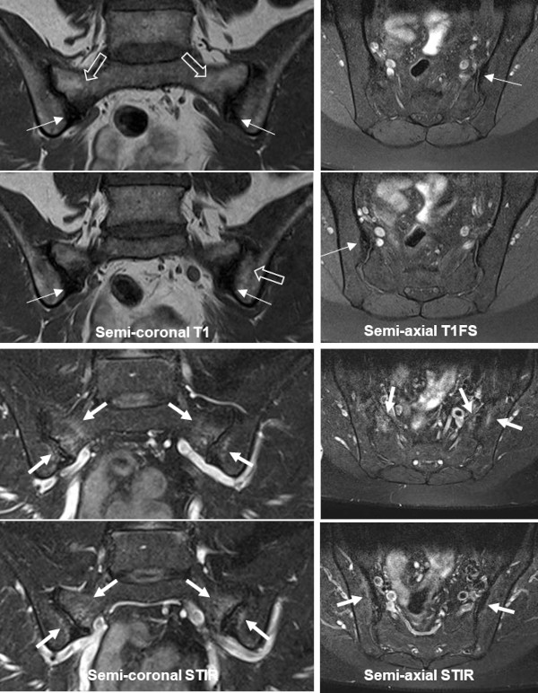
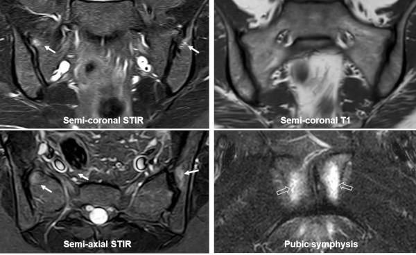
OCI is originally defined based on its radiographic appearance with unilateral or bilateral triangular-shaped sclerosis in the ileum corresponding to the weight-bearing portion of the SIJ, spared SI joint space and no evidence of erosions. OCI can be an asymptomatic incidental finding, but can also present with low back or SIJ pain. It occurs predominantly in females, especially postpartum women, but is occasionally diagnosed in men and nulliparous women where it is often related to obesity or excessive physical load to the SIJ.

Characteristically there are normal inflammatory biomarkers and HLA B27 negativity.

The pathophysiology of OCI is not fully elucidated, but previous pregnancy-related strain to the SIJ often seems to play a role, probably combined with hormonal changes.

OCI – radiography

Radiograph of the SIJ showing typical OCI changes in the form of bilateral triangular-iliac sclerosis corresponding to the load-related portion of the SIJ with preserved joint spaces.

 
OCI - MRI

The MRI appearance of OCI is characterized by manifest subchondral iliac sclerosis displaying low signal intensity on all sequences, usually located anteriorly.

There is often concomitant BME, reported present in 48-93% of individuals with OCI (doi:10.1111/1756-185X.13125). The iliac BME in OCI usually has a characteristic continuous distribution and is located peripherally to the subchondral sclerosis whereas BME as part of axSpA changes is usually discontinuous, extending to other parts of the joint outside the load-bearing areas. However, sacral BME is also frequent in patients with OCI, often located to the strain-related areas anteriorly. The BME as part of OCI may both display high signal intensity on STIR/T2FS sequences and have a depth above 1 cm, which are features often seen in active axSpA sacroiliitis. In addition, OCI changes may be accompanied by low back pain and can therefore also clinically be difficult to distinguish from axSpA. Only the manifest sclerosis, absence of definite erosion and especially the anterior location of BME changes are valid in the differentiation from sacroiliitis (doi:10.1093/rheumatology/keaa175)

In general, OCI is the most probable diagnosis in cases of recent childbirth, when relatively preserved joints margins are evident, periarticular changes are confined to the anterior portion of the SIJ and no other symptoms or signs of axSpA are present such as uveitis, inflammatory bowel disease, arthritis, dactylitis, or psoriasis.

MRI in the female shown on the radiography above complaining of low back pain, semi-coronal T1 and STIR in addition to semi-axial STIR and T1FS images. There is manifest bilateral iliac sclerosis in the anterior portion of the joints (slim arrows) with adjacent fat deposition in the sacrum and left ileum (open arrows on T1). There is a brim of BME peripheral to the iliac sclerosis in addition to anteriorly in the sacrum on both sides (arrows).

 
Pregnancy-related changes

Pregnancy-related BME is frequent and has been observed to occur both with and without pain. The occurrence of BME seems to start already during pregnancy with an increased prevalence three months after childbirth followed by a gradual decrease. At 12-month postpartum, subchondral BME has been found to occur in approx. 40% of the women sometimes accompanied by erosion-like lesions, sclerosis and fat deposition. The extent of BME may be as pronounced as in axSpA, occasionally with both intensity and depth >1 cm. This implies a risk of establishing an incorrect axSpA diagnosis in patients with low back pain at least until 12 months postpartum and therefore constitutes an important differential diagnosis to axSpA changes (doi: 10.1002/art.42457). However, BME is usually detected in the strain-related area anteriorly, especially the middle anterior portion of the joint and concomitant BME at the pubic symphysis is frequent.

Pregnancy-related changes can evolve into OCI with postpartum sclerosis and BME occurring anteriorly in the ileum, the location typical of OCI changes, as well as in the sacrum.

Postpartum changes, MRI of a woman 6 months after a normal vaginal childbirth, semi-coronal STIR and T1 in addition to semi-axial STIR image of the SIJs and a coronal STIR image of the symphysis showing BME located anteriorly at the upper part of the SIJ (arrows) with concomitant subchondral edema at the pubic symphysis (open arrows).

 
Visitors since May 1st., 2008:  1765302